

What is Orthopedic Surgery?
This specialty focuses on diagnosing, treating, rehabilitating, and preventing diseases and injuries within the musculoskeletal system of the body. Orthopedic surgeons also treat and repair bones, ligaments, tendons and joints. Doctors can conduct both non-operative treatments at their office, and operative treatments at an emergency room.
Illustrated Glossary
I. Surgical Equipment

Image 1. Bone Cutting Forceps
The Bone Cutting Forceps are used to manipulate, extract, and grasp either high-density bones or small bones. They can come either in an angled form or a straight form depending on the bone it will be utilized upon.
Image 2. Bone Holding Forceps
Bone Holding Forceps are general-purpose instruments used in medical procedures that involve bone stabilization. Bone holding forceps are designed to treat fragile bones such as smaller phalangeal and metacarpal bones requiring small instruments for circumference. An example of a bone holding forcep is the Bishop Bone Forcep, which is used to clamp bone pieces during surgical procedures. This forceps main feature is its pointed tips which allow fo better bone grasping.
Image 3. Bone Files
The Bone Files are used to carve bones. Its rounded design allows for effective maneuvering between bone and soft tissues. There are different types of bone files; for example, the double ended bone file which is used to excavate small spicules of bones from narrow spaces. Additionally, the Miler Colburn bone file is used to shaper uneven bones while the Polokoff bone file is used for smoothing the edges of bones after they have been cut.


Image 4. Bone Chisel
The Bone Chisel is used for cutting and reshaping bones in orthopedic surgeries. This tool is essential for carving into the bones in order to reach the desired surgical site. There are four types of bone chisels used: the Alexander Chisel, the Cottle Septum Chisel, the D'Errico Lamina Chisel and the Harmon Chisel. For example, the D'Errico Lamina chisel is used for cutting soft tissues during surgery.
Image 5. Bone Impactor
The Bone Impactor is used to tap bone grafts and bone parts, or fragments, into place with less trauma. This tool is also helpful for the insertion of implant devices; like for example, acetabular cups.
Image 6. Bone Rasps
The Bone Rasp is used to scrape or file away bones during surgeries. Its design allows to file bone edges without damaging the nearby tissues. The bayonet-style of the bone raps has one smooth side and one curved side with cross-serrations, which make it more suitable for filing uneven bone edges.


Image 7. Bone Burs
The Bone Burs are used to reshape and excavate different fragments of bones during surgical procedures. An example of a bone bur is the Shannon Bone Bur which is used for excavating, perforating, and cutting bone fragments.
Image 8. Bone Hooks
The Bone Hook is primarily used in procedures that involve holding and adjusting bones. This tool is preferred due to its exact precision and extremely high quality. An example of a bone hook is the Proximal Femoral Elevation Bone Hook, which is utilized to elevate the proximal femoral bone during operations.
Image 9. Hand Held Retractors
The Hand Held Retractors are utilized to hold back organs and tissue during procedures. Some of the most commonly used hand held retractors are the Capsule Retractors or Cobra Retractors with Hand Rests. Particularly, the Cobra Retractors are used to assist in hip surgical procedures.
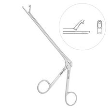


Image 10. Grasping Forceps
The Grasping Forceps are utilized to either hold or grasp tissues or organs during multiple orthopedic surgeries. For the majority of operations, this instrument is used to hold skin and tissues. Some examples of these graspng forceps include: the Grasper Rongeur and the Hoen Grasping Forceps. For example, the Grasper Rongeur is utilized for holding the rongeur tissues securely.
Image 11. Extractors
The Extractor is used to removes wires, bolts, broken pins, and threaded bolts during or after orthopedic operations. An example of an extractor is the Femoral Head Extractor which is designed to remove the femoral head during hip fracture surgeries.
Image 12. Depth Gauge
The Depth Gauge is used for measuring the depth below the surface where the surgery will be conducted. There are different sizes for the depth gauges; such as, 30mm, 50mm, 100mm and 120mm. Primarily, this instrument is used to measure, or gauge, the depth of the holes drilled into bones.


Image 13. Gouges
The Gouge is used in surgical procedures for bone contouring, or for reconstructing defects in the bone's natural form. This instrument is composed of a cylindrical blade with a bevel on either the convex or concave side. An example of a gouge is the Alexander Gouge, which is used for removing or scooping a particular part of a bone during surgery.
Image 14. Femoral Ligament Cutter
The Femoral Ligament Cutters are used to cut the femoral ligament during hip fracture or hip replacement surgeries. This instruments has different features for such as: hatt spoon tips, a fiber handle, and a length of 9 ½ inches. Particularly, the hatt spoon feature is used for femoral head and neck excision arthroplasty surgeries.
Image 15. Skin Hooks
The Skin Hooks are used to retract the skin and soft tissues during the majority of orthopedic surgeries. There are many skin hooks such as the Converse Skin Hook, Cottle Skin Hook, Freer Skin Hook, and Gillies Hook. For example, the Freer Skin Hook is used to grasp and position the skin to get a clear view of the underlying tissues, while the Gillies Hook is used to grasp, hold and/or elevate skin while closing a wound or performing reconstructive surgery.


Image 16. Bone Tamps
The Bone Tamp is used to wedge bone grafts into place. Specifically, this instrument is placed into the vertebral canal to protect the bone pieces until they can be securely put into place. An example of a bone tamp is the Bone Tamp with a Cross Serrated End, which is used to tamp wires during fracture procedures.
Image 17. Wire Passer
The Wire Passer is used for passing wires to and through the fracture sites. This instruments ergonomic design allows for a firm grip and high precision. There are different types of Passers; such as, Wire Passers, Grafts Passers, and Suture Wire Passers.
Image 18. Wire Twister
The Wire Twister is used for removing pins and wires during surgeries. During these procedures, Orthopedists use wire twisters to twist Kirschner Wires after the fixation process. Examples of wire twisters include the Corwin Wire Twister and the Jet Wire Twister. This instrument comes in a variety of shapes, sizes and lengths, while also being made of a tungsten carbide material which adds durability to the instrument.


Image 19. Kirschner Wires
The Kirschner Wires are primarily used to repair fractures. These wires, or pins, are used to hold broken bones together; however, they are required to be removed as soon as the fracture is healed. Moreover, this instrument is made of medical-grade stainless steel, which keeps them lightweight and rust-free. Lastly, these wires can be easily inserted and removed from a patient's body.
Image 20. Surgical Pliers
The Surgical Pliers are used for cutting, crimping wires, bending, removing pins and manipulating tissue during surgeries. Examples of different surgical pliers include: Flat Nose Pliers, Needle Nose Pliers, Cerclage Pliers, Wire Bending Plier, and Pin Extraction Pliers.
Image 21. Wire and Pin Management
The Wire and Pin Management surgical instruments are used in surgery to pull, bend, twist or tighten wires, pins, rods, and plates. Some of the wire and pin management instruments include the Bending Iron, Bending Pliers, Boehler Wire and Pin Tractor, and Cable Cutter. For example, the bending iron is used in spinal operations to contour rods, while the cable cutter is used for removing pins and wires.


Image 22. Surgical Screwdriver
The Surgical Screwdriver is utilized for securing surgical screws into plates when a fractured bone needs to get repaired. Additionally, these instruments attach implants to bones. There are different surgical screwdrivers, which include: the Hexagonal Phenolic Handle Screwdriver, the Cruciform Screwdriver and the Screwdriver 20mm. The hexagonal phenolic handle screwdriver is the most commonly used, and its functions include adding crews in the required depth of any surgical procedure, and minimizing hand fatigue.
Image 23. Phenolic Handle Mallet
The Surgical Mallets are instruments utilized for the manipulation, cutting, and scraping of the bones during a surgical procedure. The most commonly used surgical mallet for orthopaedic surgeries is the Phenolic Handle Mallet, which is used to manipulate bones and cut, split and scrape the bones being operated on.
Image 24. Socket Wrench
The Socket Wrench is a surgical tool which is the combination of ratchet and socket. Its function includes being used for tightening or loosening pins or wires durong orthopaedic procedures. In addition, the socket wrench has the following features: a T-shaped handle, a 11mm tip end and an overall length of 7".


Image 25. Spiked Disc
The Spiked Disc is an instrument ideal for internal fractures fixation during orthopaedic procedures. The spiked disc has the following features: has a diameter of 25", is rustproof and is lightweight. This tool can be cleaned and sterilized easily and it is reusable. The body of the spiked disc is also roust and is classified as unbendable. Moreover, its round shape is the reason why it is so commonly utilized in hip and knee surgeries.
Image 26. Drill Guide
The Drill Guide, also known as Drill Sleeve, is an instrument designed to create holes of specific sizes during orthopaedic surgeries at fracture sites. There are a few types of Drill Guides, or Sleeves, which include: the Double Drill Guide, the Drill Bit, the Drill Guide Neutral, the Insert Drill Sleeve.
Image 27. Poole Suction Tubes
The Poole Suction Tubes are a very versatile surgical instrument because its function. The primary function of the suction tube is to assist in evacuating large quantities of pooled blood, debris, and fluid within the surgical site. This instrument is used for the majority of operations.


Image 28. Osteotomes
The Osteotome is an instrument utilized for preparing and manipulating bones during surgery. An osteotome is similar to another commonly used instrument, the Chisel, but it they differ because the osteotome is two-sided beveled. An commonly used osteotome is the Stubby Osteotome thats 7cm long. The blunt blade from this osteotome makes it easier to conduct delicate work in confined spaces between bones.
Image 29. Punches
The Punches are instruments utilized for removing either small or large portions of tissues and/or bones during orthopaedic surgeries, especially in the biopsy. Some examples of punches include: Keyes Punches, Micro France Punches, and Meniscal Crescent Punches.
Image 30. Measuring Instruments
The Measuring Instruments are utilized to measure a variety of bones and muscles during surgical procedures. Popular measuring tool patterns include: the Flexible Ruler, the Castroviejo Caliper, the Steel Ruler, the K-Wire Ruler and Pin Gauge, and the 8" Bone Compass. Specifically, the 8" Bone Compass is utilized to make accurate measurements of the patient and perform error-free incisions. In addition, these instruments are reusable and do not malfunction easily.


Image 31. Acetaminophen (Tylenol)
The medication Acetaminophen is utilized to relieve mild and/or moderate pain from headaches, back pain and muscle aches, and reduce fever that might commence after an orthopaedic surgery. However, acetaminophen does not reduce inflammation. These types of medication maybe come in the form of chewable tablets, capsules, suspensions, and orally disintegrating tablets, also known as “meltaways.” One of the most common tablets with acetaminophen as its active ingredient is Tylenol.
Image 32. Aspirin (Excedrin)
The medication Aspirin is commonly used in orthopaedic practices to treat and/or manage many forms of arthritis. However, aspirin is only a part of a total treatment program for arthritis, meaning it Is not a cure for arthritis. The primary function of Aspirin is to reduce the effects of inflammation post-surgery, a reaction of the body that causes pain, swelling, redness, and heat in patients.
Image 33. Ibuprofen (Advil)
The medication Ibuprofen is utilized for managing moderate to severe pain aches after orthopaedic surgery. This medication reduced both soreness, swelling and stiffness in joints, and can even be combined with opioids for stronger effects. Specifically, intravenous, or IV, ibuprofen is utilized to relieve acute pain post-surgery and minimize the opioid use of a patient in the first 48 hours after an orthopaedic trauma.


Image 34. Corticosteroids
The medication known as Corticosteroids is a steroid utilized for relieving the pain of inflammation and soreness located in one area. An injection of corticosteroids into a joint, such as a shoulder or knee joint, can relieve some of the pain felt by the patient for typically four to six months. The primary conditions treated with corticosteroids are athletic injuries, nerve compression and osteoarthritis. Nevertheless, corticosteroid-use can cause the development of infections, or make them get worse. In addition, corticosteroid-use raises the level of sugar in the blood; thus, diabetes patients must be very careful when using this type of medication.
Image 35. Lidocaine
Local anesthetics’ primary function is to block pain in a small area of the body, typically in the area where there has been orthopaedic trauma, by blocking the pain signals from the nerves to the brain. Local anesthetics are used as anesthesia during a procedure, or as part of a pain management program after surgery. The most commonly used local anesthetics include lidocaine, bupivacaine, and ropivacaine. However, it is important to make note that using local anesthetics may cause a possible allergic reaction in a patient, which can lead to side effects like nerve damage, muscle spasms, and convulsions. Nevertheless, these side effects can be avoided when a patient provides a through and complete medical history.
Image 36. Diazepam (Valium)
Muscle relaxants’ primary function is to have an overall sedative, or numbing effect, on the body. These relaxants are typically prescribed before orthopaedic surgery and are usually taken for a short-term basis. Examples of commonly used muscle relaxants are: Carisoprodol (Soma), Cyclobenzaprine (Flexeril), and Diazepam (Valium). While muscle relaxants are beneficial, since they work as a pre-surgery treatment, some muscle relaxants may be habit-forming and addictive, meaning patients must be aware of this medication usage.


Image 37. Calcitonin (Miacalcin)
The medication known as Osteoporosis drugs have been recently approved to treat osteoporosis and reduce the risk of fractures in older patients. Since osteoporosis is a condition that causes bones to thin, which can lead to very painful bone fractures, several drugs have been appointed for this type of treatment. Osteoporosis drug’s primary function is to reduce the loss of bone density and increase the amount of calcium that is being deposited into the bones. Examples of commonly used osteoporosis drugs are: Calcitonin (Miacalcin) and Alendronate (Fosamax). Specifically, Calcitonin, the drug seen in the picture, is a hormone that enhances bone-forming cells.
Image 38. Pain-Relieving Rubs (Bengay)
Analgesic, or pain-relieving, rubs are ointments that contain some form of salicylate. Analgesic rubs are a form of pre-surgery treatment that helps with for short-term pain relief. To apply analgesic rubs, a patient must rub the ointment on their skin over the area where there is the most pain. This cream then produces a numb or warmth-like sensation over the affected area which may provide some sort of relief from the pain. However, these ointments do not reduce inflammation in the area.
Image 39. Trolamine Salicylate Cream
Trolamine Salicylate is an analgesic ointment that comes in the form of a skin cream, that primarily treats minor joint pain and muscle aches, while also reducing inflammation. This cream must be applied at intervals and around three-to-four times per day. The conditions most commonly treated with this cream are arthritis, back pains and sprains. An example of an ointment with 10% Trolamine Salicylate is Aspercreme. Lastly, this ointment also belongs to the class of drugs classified as salicylates.
Most Common Procedures


Photo Credit: Verónica Bado

Image 40. Cortisone Injections
As mentioned previously, the primary use of cortisone injections are for relieving the pain of inflammation, irritation and soreness located in one area. The joints most commonly injected with corticosteroids include: elbow joint, knee joint and shoulder joint. Nevertheless, small joints in the fingers and the feet also benefit from these injections. The most common area injected with corticosteroids, seen throughout my shadowing, was the shoulder joints.
Image 41. Cast Installations and Removals
A cast is an orthopaedic instrument that holds a fractured bone in place while it heals. For a bone to properly heal, it must be immobilized; thus, casts are utilized as effective methods of immobilization and to prevent an increase or decrease of muscle contractions. Specifically, casts immobilize the joint above and below the area that has been fractured that needs to be kept straight and without motion. Moreover, casts are primarily made of plaster and fiberglass, which is the colored part of the cast, but it also includes cotton to make the cast soft and padded. While wearing a cast, people might need medical instruments like crutches, walkers and wheelchairs to move around, depending on the severity of the fracture. Casts also must be kept clean and dry. Lastly, casts may stay on a patient for four to 12 weeks.
Image 42. Splints
A splint is an orthopaedic instrument that stabilizes an injured area to reduce its movement either temporarily or until it heals completely. Additionally, a splint might be utilized on a fractured bone or on an area with a torn ligament or a severe laceration. Depending on the location and severity of the injury, a splint requires a specific shape, size, length and material. Particularly, a splint is made of rigid metal or plastic, which support, protect and immobilize the injured area, but not as effectively as a cast. This is because splints require a high patient compliance and responsibility to use the splint adequately.
I. In the Office
II. In the Hospital
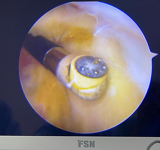
Photo Credit: Verónica Bado

Photo Credit: Verónica Bado
Image 43. Arthroscopies
Arthroscopy is a surgical procedure utilized for treating joint problems. This procedure consists of inserting a narrow tube, which is attached to a fiber-optic video camera, through a small incision made in the affected area. The camera then transmits a high-resolution video of inside the affected joint to a high-definition video monitor. The advantage of an arthroscopy is that it allows the surgeon to see inside of the joint without making a large incision. Moreover, surgeons can even repair some types of joint damage during an arthroscopy, with pencil-thin surgical instruments inserted through additional small incisions near the original incision. Some surgical procedures treated with arthroscopies include: removing loose bone fragments, fixing torn ligaments, and viewing inflamed joint linings. Furthermore, arthroscopies are used to diagnose a variety of conditions which affect primarily areas like the knee, shoulder, ankle, hip and wrist.
Image 44. Repairing Bone Fractures - Ankle Fracture
An ankle fracture is the 4th most common fracture in adults; however, ankle fractures can occur at any age. Typically, ankle fractures result from twisting or rolling your ankle while running or walking, or due to falls from big heights like a ladder. Moreover, a fractured ankle can either be a single broken bone or several broken bones, which can affect a person’s ability to walk. Lastly, ankle fractures can be classified depending on the number of pieces that have broken away from their original position. As a result, ankle fractures can be classified as nondisplaced fractures, displaced fractures or open fractures.
Image 45. Repairing Bone Fractures - Wrist Fracture
The wrist is the joint at the end of the forearm, and its this hinge joint between the arm and the hand that allows a person to reposition their hand. In total, the wrist possesses eight bones that are arranged into two rows, the proximal and the distal rows. Specifically, a wrist fracture occurs when a person has fractured one of the smaller, carpel bones, or the distal radius, which is the larger of the two bones that make up the forearm. The distal radius typically fractures near the wrist and the bones of the thumb. The two groups that most commonly have wrist fractures are elderly people, due to the loss of bone density, and athletic young people, who fracture these bones during rigorous exercises. Wrist fractures can be healed with either a splint of a cast, depending on the severity of the fracture; however, patients need physical therapy in order to recuperate strength and mobility. If the fracture needs surgery, these fractures are usually fixed with metal plates and pins that hold the bone in place.


Image 46. Osteoarthritis
Arthritis is a common illness in the orthopaedic world that is defined as the inflammation of a joint which is typically accompanied by pain and swelling. However, Osteoarthritis is a degenerative type of arthritis which is characterized by the degradation of cartilage over time, due to a lifetime of use of due to a traumatic injury. Throughout the progression of this disease, the underlying bone becomes exposed, which causes the joint to become more painful to move; and thus, a person’s range of motion becomes heavily diminished. With osteoarthritis, pain is typically worse activity but alleviates with rest. Lastly, this type of arthritis is treated with anti-inflammatory medication like Aspirin.
Image 47. Post-Traumatic Arthritis
Post-Traumatic Arthritis is a type of Osteoarthritis that is caused by a bone fracture and/or a dislocation. This type of illness typically occurs after an injury to the joints and is accompanied by symptoms like stiffness and moderate pain. For this type of arthritis, patients usually do not need to undergo surgery; however, it might take a few months to feel better. Lastly, post-traumatic arthritis is seen more commonly in younger people, like teens and kids, than in older adults or the elderly.
Image 48. Rheumatoid Arthritis
Rheumatoid Arthritis is an autoimmune, systemic and inflammatory illness that affects primarily the joints, and usually, many joints all at once. When a joint is afflicted with rheumatoid arthritis, the lining of the joint, the synovium, becomes inflamed, thus causing damage to the joint tissue. The result from this damage can be chronic pain, a lack of balance, and even deformities in those joints. The most commonly affected joint includes the hands, knees and wrists, elbows and ankles.


Image 49. Carpal Tunnel Syndrome
Carpal Tunnel Syndrome is an illness where the median nerve, which originates from the spinal cord and extends to the fingers, is compressed or squeezed as it passes through the carpal tunnel in the wrist, which is a narrow, confined space. Since the median nerve is a sensory and motor nerve of the arm, symptoms from this illness include feeling numbness in the fingers, pain in the hand and wrist, and the fingers becoming useless.
Image 50. Lateral Epicondylitis - "Tennis Elbow"
Lateral Epicondylitis, also known as “Tennis Elbow,” is caused by the damage suffered by the tendons that bend the wrist backward away from the palm. The tendon associated with lateral epicondylitis is called the extensor carpi radialis. The swelling of these tendons causes symptoms like pain and swelling.
Image 51. Medial Epicondylitis - “Baseball Elbow"
Medial Epicondylitis, also known as “Baseball Elbow,” is caused by the damage suffered by the tendons that bend the wrist toward the palm. Specifically, the side affected by this illness is the medial side of the arm. The tendon associated with lateral epicondylitis is called the flexor carpi radialis.


Image 52. Osteoporosis
Osteoporosis is a bone disease that develops when the bone is not replaced as fast as it is removed. Specifically, it develops when the bone mineral density and overall bone mass decrease, thus changing the structure of the bone with time. This harmful change leads to a decrease in bone strength, which in turn, increases the risk of bone fractures. Three common causes of osteoporosis include a decrease in hormones like estrogen and testosterone, prolonged bed-ridden illnesses, and medical conditions which cause increased inflammation throughout the body.
Image 53. Scoliosis
Scoliosis is a condition which presents itself as a lateral curvature of the vertebrae, or spine, of a person, thus giving the appearance that the person is leaning more to one side than the other. Individuals that suffer from scoliosis typically have an S-shaped or C-shaped curve of the spine. While there is no cure for scoliosis, there are treatments to deal with this condition which include surgeries to align the spine and reduce the severe angle of the curvature. Lastly, scoliosis is a condition that can affect anyone, and at any age.
Image 54. Tendonitis
Tendonitis is a condition where the tendons, or tendon coverings, between the muscles and bones in any area of the body become inflamed. This condition usually happens due to the repetitive use and strain of an area. Tendonitis can develop in the shoulder, hips and elbows; however, rest from high-energy activities helps the affected tendons heal. A type of tendonitis is called “Tennis Elbow,” also known as Lateral Epicondylitis, which was mentioned previously.
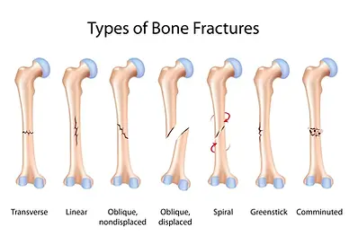

Image 55. Trigger Finger
Trigger Finger is an illness that is caused by an irritation of the digital sheath surrounding the flexor tendons of the fingers. This illness is triggered when the tendon sheath becomes swollen and it pinches the tendon, which prevents the tendon from gliding and moving smoothly. The name “Trigger Finger” comes from when the tendon pinches itself and the suddenly releases and though a trigger were being released.
Image 56. Bone Fracture
A bone fracture, or break, is a condition characterized by a crack or fissure in the bone. A bone fracture typically occurs when a force greater than the force a bone can withstand is exerted upon said bone, which then proceeds to crack. While the most common places for bones to fracture are the ankle, wrist and hip, bones can fracture in other places such as: fingers, toes, knees, femurs, tibia, fibula, radius, elbow, clavicle, and many more.
Image 57. Avascular Necrosis (AVN)
Avascular Necrosis, also called AVN, is a bone tissue illness that is caused by an injury or previous disease which results in a disrupted blood supply in an area of the body. This impaired blood supply causes symptoms like severe pain and can cause a weakened bone to collapse. Particularly, if avascular necrosis is developed near a joint, it is possible that the surface of the joint may collapse.


Image 58. DeQuervain’s Syndrome
DeQuervain’s Syndrome is a condition located at the wrist, on the side closest to the thumb, which occurs because tendons become trapped beneath a ligament as they traverse toward the thumb. Specifically, the tunnel where these tendons travel through is more narrow than usual due to the thickening of the soft tissues that make up said tunnel. As a result, hand and thumb motions cause incredible pain, specifically when holding, grasping or twisting something. This condition can be treated with a splint, a cortisone injection in the affected areas or surgery in order to relieve the pressure of the ligament on the tendon.
Image 59. Herniated Disc
A Herniated Disc is a condition caused by the rupture, or slipping, of the outer cartilage section of a disc. This is caused because the cushions that are located between the disc in the vertebrae are pushed outside of their normal, resting position. Symptoms from herniated discs include sharp pain in one side of the body, weakness in that one side of the body, and numbness in the legs or arms.
Image 60. Plantar Fasciitis
Plantar Fasciitis is an overuse illness that is caused by the inflammation of the tough, fibrous tissue that connects the heel to the base of the toes. This illness is caused primarily by foot structure, the type of shoes used, and excessive walking and foot movement. The main symptom of plantar fasciitis is moderate heel pain; however, if left untreated, this pain may radiate to other areas such as the hips, back and knees.
Anatomical & Histological Concepts


Image 61. Subchondral Tissues
Subchondral Tissues are the tissues located at the very ends of the bones, which are covered by cartilage.
Image 62. Compact Tissues
Compact Tissues are the harder, outer tissues of bones.
Image 63. Cancellous Tissues
Cancellous Tissue are the sponge-like, inner tissues of bones.


Image 64. Cartilage
Cartilage is a specialized type of tissue that connects joints with one another, and cushions the bone, thus allowing the joints to move without pain.
Image 65. Ligaments
Ligaments are classified as bands of different types of tissue whose primary function is to connect bones, joints and organs and hold them in their place.
Image 66. Periosteum
The periosteum is a thin yet tough outer membrane that covers the bones; and typically, it is attached to muscles, ligaments and/or tendons.
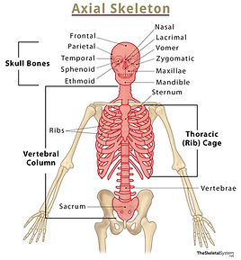
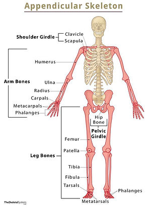

Image 67. Axial Bones
Some of the most common axial bones are the head, ribs and sternum. These make up for 80 out of the 206 bones in the body.
Image 68. Appendicular Bones
Some of the most common appendicular bones are: arms, shoulders, wrists, hands, legs, hips, ankles and feet. These make up for 126 out of the 206 bones in the body and are the most commonly fractured bones.
Image 69. Osteoblasts
Osteoblasts are cells located within the bones. The osteoblasts’ primary function is to form new bone tissues.


Image 70. Osteocytes
Osteocytes are cells located within the bones. The osteocyte’s primary function is to maintain the bone as a living tissue.
Image 71. Osteoclasts
Osteoclasts are large cells located and formed within the bone marrow. The osteoclast’s primary function is to absorb and remove any unwanted tissue present.
Image 72. Hematopoietic Cells
Hematopoietic cells are cells located within the bone marrow which are responsible for producing red blood cells, white blood cells and platelets.


Image 73. Cartilaginous Joint
A Cartilaginous Joint, as the name suggests, is a joint where adjacent bones are bound together by cartilage. While these types of joints lack a joint cavity, they involve bones that are joined together by hyaline cartilage or fibrocartilage. Moreover, there are two types of cartilaginous joints called the synchondrosis, which is a joint joined by hyaline cartilage, and the symphysis, a joint joined by fibrocartilage. An example of a cartilaginous joint is the pubic symphysis.
Image 74. Hinge Joint
A Hinge Joint is a joint characterized by its movement of opening and closing in one single direction. In similarly to ball and socket joints, a hinge joint can also be affected by osteoarthritis, an illness mentioned previously. In addition, hinge joints are more commonly dislocated due to sport injuries. Some examples of hinge joints include: fingers, toes, elbows and knees.
Image 75. Condyloid Joint
A Condyloid Joint, also known as an Ellipsoid Joint, is a joint composed of bone called a condyle, which fits into an egg shape-like cavity. Specifically, condyloid joints only allow forward and backward, and side-to-side movement, but it does not allow rotational movement. Common illnesses that affect condyloid joints include arthritis and Carpal Tunnel Syndrome. An example of a condyloid joint is the fingers.


Image 76. Saddle Joint
A Saddle Joint is a joint characterized by being made up of two bones, one which is concave and another that is convex. These two bones resemble a saddle and a rider, respectively. Similarly, to ball and socket joints and hinge joints, a saddle joint can also be affected by osteoarthritis. Some examples of saddle joints include: the shoulder and the thumb.
Image 77. Pivot Joint
A Pivot Joint, also known as Rotary Joints, are joints which are composed of two bones, one bone that moves within a ring-like structure, and second bone that allows rotation. A pivot joint can also become affected by arthritis of common wear-and-tear problems. An example of where a pivot joint is located is with the ulna and radius.
Image 78. Ball and Socket Joint
The most commonly known joint is called a Ball and Socket Joint. This type of joint is also composed of two bones, where one bone possesses a rounded head which fits into a cup-like, hollow space of another bone. The ball and socket joints allow for movement in all directions, due to the ability of the rounded-head bone to move freely with the socket of the second bone. An example of a ball and socket joint is the shoulder, which can be affected by osteoarthritis.

Orthopaedic Surgeon
A physician who's work composes of diagnosing, treating and managing the rehabilitation process for patients who suffer from either injury or disease in any area of the musculoskeletal system.
Musculoskeletal System
The Musculoskeletal System is classified as a complex system which includes the body’s muscles and skeleton, and the joints, ligaments, tendons, and nerves.
R.I.C.E
Acronym for Rest, Ice, Compression and Elevation.
Technical Vocabulary

Electromyogram (EMG)
An Electromyogram, also called EMG, is a test which evaluates the nerve and muscle functions of an individual.
Orthopaedic Emergencies
Orthopaedic Emergencies occur when a person injures a bone or a soft tissue in such a way that it requires immediate medical attention to avoid harm to the body. These emergencies deal with sudden and acute injury or trauma, or they could be complications from existing condition, illness or surgery. The most common orthopaedic emergency surgery is bone fracture surgeries, which require bone alignment and either a splint or a cast.
Post-Surgery Trauma
Post-surgical trauma is characterized as the distressing emotional and physical impact that a surgery has on a patient’s mind and body. This disorder can embark a variety of causes and can also have a varied range of consequences. This type of trauma has more psychological effects on a patient.